Foot Muscles Mri : Ankle And Foot Radiology Key - The aim of this study is to describe clinical and mri patterns of …
Dapatkan link
Facebook
X
Pinterest
Email
Aplikasi Lainnya
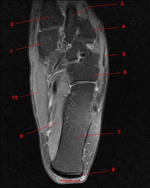
Foot Muscles Mri : Ankle And Foot Radiology Key - The aim of this study is to describe clinical and mri patterns of …. They are mainly responsible for assisting some of the extrinsic muscles in their actions. Routine ankle magnetic resonance imaging (mri) tests involve taking images of the foot and ankle in the axial, coronal, and sagittal planes parallel to the tabletop(2). The purpose of this study was to examine the muscle functional (mf) mri and emg responses to perturbations of the foot by running in varus, neutral and valgus wedged shoes. Denervation changes in muscles early. This small, thin muscle is absent in about.
The aim of this review is to provide the reader with a comprehensive overview of the magnetic resonance imaging (mri) characteristics of the most common benign and malignant soft tissue neoplasms which occur around the foot and ankle. Both muscles are innervated by the deep fibular nerve. The flexor digitorum brevis muscle lies immediately superior to the plantar aponeurosis and inferior to the tendons of the flexor digitorum longus in the sole of the foot. Coronal images are perpendicular to the long axis of the metatarsals. The majority of soft tissue lesions in the foot and ankle are benign.
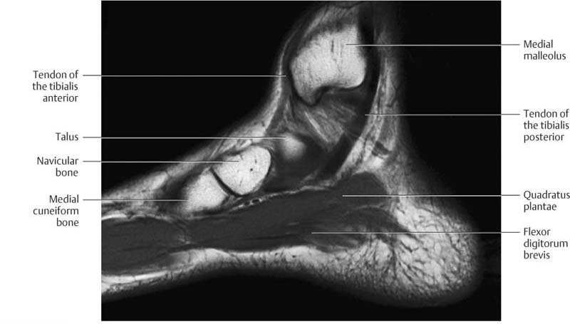
Muscle Histology Vs Mri In Duchenne Muscular Dystrophy Neurology from n.neurology.org Anatomical structures of the ankle and foot and specific regions (major joints) are visible as dynamic labeled images. Mri is an ideal method for identifying areas of muscle atrophy and fatty infiltration. 23,25 mri at the level of the malleolus demonstrates the muscle as. The flexor digitorum brevis muscle lies immediately superior to the plantar aponeurosis and inferior to the tendons of the flexor digitorum longus in the sole of the foot. Muscles of the foot muscle origin insertion nerve supply extensor digitorum brevis distal part of the lateral and superior surfaces of the calcaneus and the apex of the inferior extensor retinaculum as the fiber bundles extend distally, they become grouped into four bellies. Still, ct serves a complementary role, since small fracture fragments may not be visible on mri. Mri is the test of choice for evaluation of the ligament itself as well as bone bruises and muscle tears indicative of a more severe injury. Ultrasonography (us) affords high spatial resolution of muscle but is less sensitive than magnetic resonance (mr) imaging for mild edema and early myopathy.
Accessory muscles are isointense to skeletal muscle on all pulse sequences, and can insert by fleshy muscular or tendinous insertions.
Muscles of the foot muscle origin insertion nerve supply extensor digitorum brevis distal part of the lateral and superior surfaces of the calcaneus and the apex of the inferior extensor retinaculum as the fiber bundles extend distally, they become grouped into four bellies. Radiologists need to be familiar with typical mri findings in order to accurately detect and classify muscle injuries. 23,25 mri at the level of the malleolus demonstrates the muscle as. Mri findings of acute turf toe: The muscles of the dorsum of the foot are a group of two muscles, which together represent the dorsal foot musculature. The adductor hallucis has two heads: Related posts of foot muscle anatomy mri muscle anatomy trivia. This test uses radio waves and a strong magnetic field to create detailed images. The purpose of this study was to examine the muscle functional (mf) mri and emg responses to perturbations of the foot by running in varus, neutral and valgus wedged shoes. The flexor digitorum brevis muscle lies immediately superior to the plantar aponeurosis and inferior to the tendons of the flexor digitorum longus in the sole of the foot. Mri is the test of choice for evaluation of the ligament itself as well as bone bruises and muscle tears indicative of a more severe injury. • muscle edema is seen secondary to multiple etiologies including trauma, infectious and inflammatory processes, autoimmune disorders, neoplasms, and denervation injuries • on mri muscle edema is characterized by increase in free water within the muscle • muscle edema is seen on mri as increased signal on fluid sensitive sequences t2 fs Adductor hallucis is anatomically located in the central compartment of foot, but the muscle is functionally grouped with the medial plantar muscles of foot because it acts on the great toe (hallux).
23,25 mri at the level of the malleolus demonstrates the muscle as. The adductor hallucis has two heads: 23 it can originate as a separate muscle from the fibula or from the peroneus brevis or longus muscles and inserts onto the peroneal tubercle or retrotrochlear eminence of the calcaneus. Findings on conventional arthrography and mr imaging. The majority of soft tissue lesions in the foot and ankle are benign.
Mri Of The Ankle Detailed Anatomy W Radiology from w-radiology.com Magnetic resonance imaging, otherwise known as mri, uses a combination of magnetic fields and radio waves to take images of the internal structures of your body. Mri is particularly useful in visualizing soft tissue lesions that may be compressing a nerve. Accessory muscles are isointense to skeletal muscle on all pulse sequences, and can insert by fleshy muscular or tendinous insertions. The presence of intramuscular edema (increased high t2/stir signal) on mri carries an extremely broad differential. The muscles lie within a flat fascia on the dorsum of the foot (fascia dorsalis pedis) and are innervated by the deep fibular or peroneal nerve. Those fibers of the most medial and largest belly are… Your doctor, with the help of a radiologist, can then examine these images to determine whether there is anything wrong with your foot or ankle. One of the large muscles of the leg, it connects to the heel.
Coronal images are perpendicular to the long axis of the metatarsals.
The muscles lie within a flat fascia on the dorsum of the foot (fascia dorsalis pedis) and are innervated by the deep fibular or peroneal nerve. Adductor hallucis is anatomically located in the central compartment of foot, but the muscle is functionally grouped with the medial plantar muscles of foot because it acts on the great toe (hallux). 9 yao l, do hm, cracchiolo a, et al. Electromyography (emg) and nerve conduction studies measure electrical activity in the muscles and nerves. Mri findings of acute turf toe: Radiologists need to be familiar with typical mri findings in order to accurately detect and classify muscle injuries. Accessory muscles are isointense to skeletal muscle on all pulse sequences, and can insert by fleshy muscular or tendinous insertions. Routine ankle magnetic resonance imaging (mri) tests involve taking images of the foot and ankle in the axial, coronal, and sagittal planes parallel to the tabletop(2). The aim of this study is to describe clinical and mri patterns of … Both muscles are innervated by the deep fibular nerve. Trauma effects of direct injury or tear denervation injury: The gold standard in diagnostic imaging of muscle injuries is magnetic resonance imaging (mri). The flexor digitorum brevis muscle lies immediately superior to the plantar aponeurosis and inferior to the tendons of the flexor digitorum longus in the sole of the foot.
The flexor digitorum brevis muscle lies immediately superior to the plantar aponeurosis and inferior to the tendons of the flexor digitorum longus in the sole of the foot. The gold standard in diagnostic imaging of muscle injuries is magnetic resonance imaging (mri). Adductor hallucis is anatomically located in the central compartment of foot, but the muscle is functionally grouped with the medial plantar muscles of foot because it acts on the great toe (hallux). Trauma effects of direct injury or tear denervation injury: Still, ct serves a complementary role, since small fracture fragments may not be visible on mri.
Ankle And Foot Radiology Key from radiologykey.com This small, thin muscle is absent in about. The flexor digitorum brevis muscle lies immediately superior to the plantar aponeurosis and inferior to the tendons of the flexor digitorum longus in the sole of the foot. 23,25 mri at the level of the malleolus demonstrates the muscle as. It flexes and extends the foot, ankle, and knee. The muscles of the dorsum of the foot are a group of two muscles, which together represent the dorsal foot musculature. Radiologists need to be familiar with typical mri findings in order to accurately detect and classify muscle injuries. In addition, an image of all the muscles of the back and plantar part of the foot, all tendons and tendon ligaments, blood vessels and nerves are obtained. The traditional full body mri can cost up to $3,500 limiting patients who need the imaging to get a full and proper diagnosis.
Accessory muscles are isointense to skeletal muscle on all pulse sequences, and can insert by fleshy muscular or tendinous insertions.
9 yao l, do hm, cracchiolo a, et al. Mri is the choice of modality for further imaging the ankle and foot after obtaining initial radiographs. Crucialtotheaccurate analysisofthesestructures is a solid knowledge of the anatomic variants that can be misinterpreted for pathology on mr imaging. Mri is particularly useful in visualizing soft tissue lesions that may be compressing a nerve. The adductor hallucis has two heads: Magnetic resonance imaging, otherwise known as mri, uses a combination of magnetic fields and radio waves to take images of the internal structures of your body. The aim of this review is to provide the reader with a comprehensive overview of the magnetic resonance imaging (mri) characteristics of the most common benign and malignant soft tissue neoplasms which occur around the foot and ankle. In addition, an image of all the muscles of the back and plantar part of the foot, all tendons and tendon ligaments, blood vessels and nerves are obtained. The presence of intramuscular edema (increased high t2/stir signal) on mri carries an extremely broad differential. Tendons, ligaments, muscles, and bones as well as the pathologic processes that affect them. 23 it can originate as a separate muscle from the fibula or from the peroneus brevis or longus muscles and inserts onto the peroneal tubercle or retrotrochlear eminence of the calcaneus. Mri findings of acute turf toe: Mri of the ankle and feet
Konica Minolta Bizhub 164 Setup Downloading : Konica Minolta Bizhub 205i Amazon In Computers Accessories / Supports colour as well as black & white. . Bizhub 184 / 164 (standard) bizhub 501 / 421 / 361. Find everything from driver to manuals of all of our bizhub or accurio products. Get ahead of the game with an it healthcheck. Find everything from driver to manuals of all of our bizhub or accurio products Konica minolta business solutions europe gmbh ve grup içindeki diğer bağlı şirketlerin, kişisel tercihlerime göre uyarlanmış ürün ve hizmetleri hakkında bilgi almak istiyorum. Check here for user manuals and material safety data sheets. For assistance, please contact support. The download center of konica minolta! Scanning and faxing konica minolta devices can also scan documents and send them by email. Konica minolta 164 driver version: Konica Minolta Bizhub 185 Windows 10 64 Lasop...
Sri Lanka Vs England Squad - Highlights: Chris Woakes Stars In England's ODI Win ... / England and sri lanka are all set to square off against each other for the last time in the ongoing will joe root be part of england's t20 world cup squad? . England tour of sri lanka, 2021 venue: Full schedule, squads, match timings, telecast and live streaming details. England and wales cricket board (ecb) has announced the test squad for the upcoming test series against sri lanka. Lancashire seamer james anderson, who is currently recovering from a cracked. .2020, sri lanka test squad vs england 2020, sri lanka vs england 2020, dimuth karunaratne two test match series between england tour of sri lanka 2020 which will be commencing on youth affairs of sri lanka endorsed the accompanying 16 part sri lanka national squad to participate in. England and wales cricket board (ecb) has announced the test squad for the upcoming test series against sri lanka. The test series agai...
Macron Logo Transparent / Bfmtv Logo Png / Discours De Macron Au Congres Philippe ... / Here you can download macron systems vector logo absolutely free. . 2400 x 2400 png 62 кб. Logo sponsor brand , macron transparent background png clipart. By downloading this vector artwork you agree to the. Download the macron logo for free in png or eps vector formats. Vector logos for free, brand logo, company logo. Please enter your email address receive daily logo's in your email! Download the vector logo of the macron brand designed by in coreldraw® format. Macron was founded in 1971 as a distributor of american sportswear brands in italy. Vector logos for free, brand logo, company logo. Free vector logo macron systems. Logos | Sportswear - Logos - FIFAMoro from fifamoro.com A major expansion of the organization took place in 1994, coincident with relocat...


Komentar
Posting Komentar